From Ars Technica · · 1m 40s
Nikon's microscopy contest celebrates motion at the cellular level
A Chinese engineer won by filming the abnormal beating of cilia in a rare genetic respiratory disease.
In brief
A Chinese engineer won by filming the abnormal beating of cilia in a rare genetic respiratory disease. Advanced microscopy that captures living cells in motion reveals disease mechanisms that still images cannot show. Originally reported by Ars Technica.
Chinese engineer wins Nikon microscopy contest
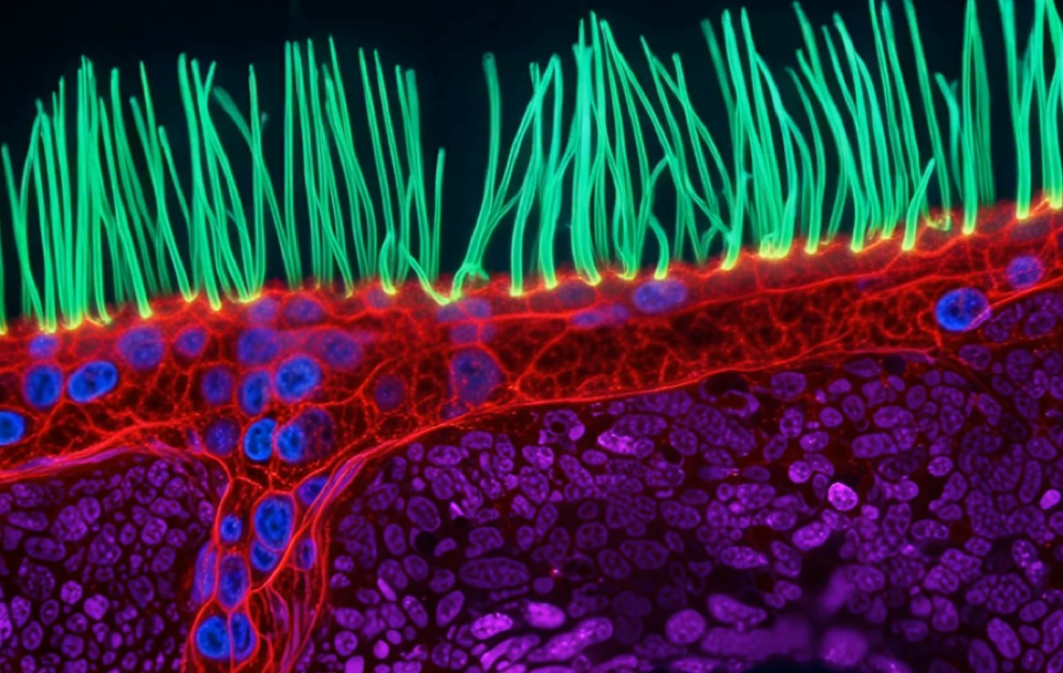
Ning Xu from Tsinghua University in Beijing beat 346 other entries from 40 countries with a super-resolution video of abnormal airway cilia in a child with a rare genetic respiratory disorder.
Nikon's Small World in Motion explained
Founded in 2011, the competition is a spinoff of Nikon's Small World Photomicrography Competition from 1974. Both showcase the beauty and complexity of microscopic structures, one through still photos and one through video.
What is primary ciliary dyskinesia
PCD is a genetic disorder where tiny hairlike structures in the lungs don't work properly. Cilia normally move germs and mucus out of the body, but in PCD they may be the wrong size, shape, or missing entirely, causing respiratory infections.
Why video is essential for studying cilia
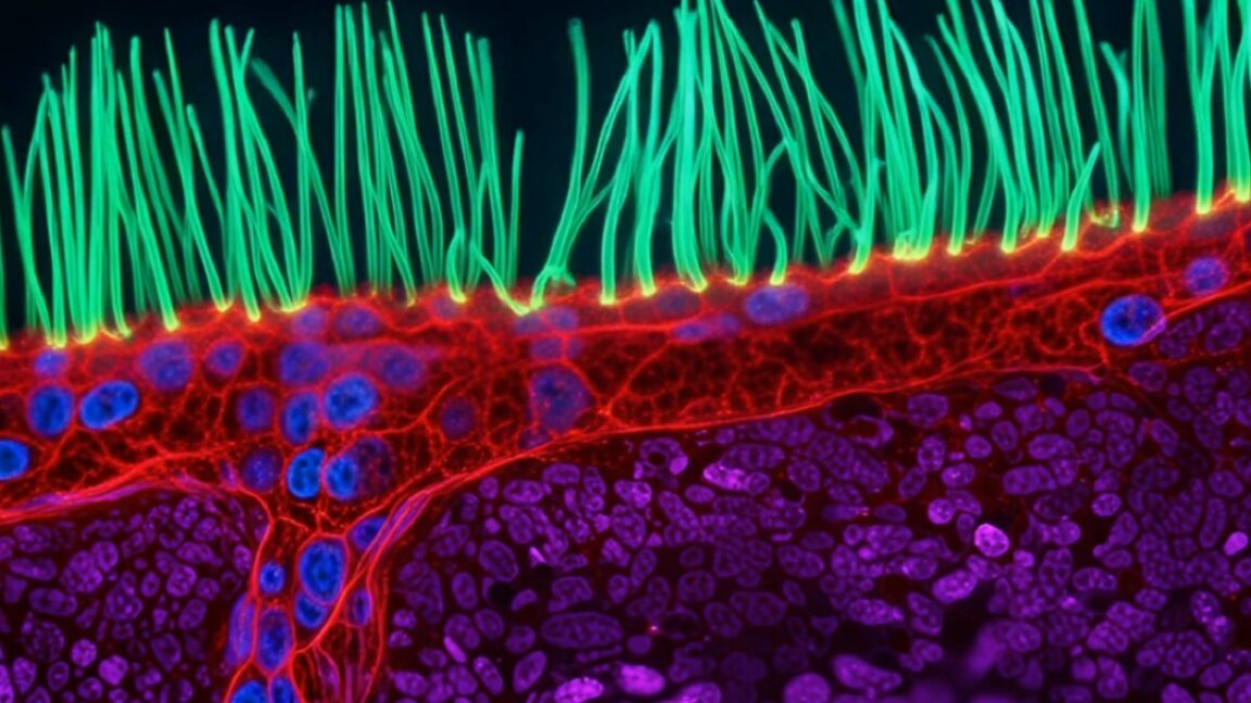
Cilia are tiny and move rapidly, making them hard to image while alive. Many advanced microscopy techniques use intense light or fluorescent dyes that would damage living samples, so researchers need methods that minimize light exposure.
“To understand this disease, you need to observe around 10 seconds of motion, not just a still image.”
Diffractive super-resolution microscopy explained
Xu used this technique, which employs multiple light waves and a digital micromirror to precisely shape light. It minimizes light reaching the living sample while capturing rapid cilia movement at high resolution.
Other winning entries in the competition
Second place went to a video of a roundworm and single-celled organism. Third place featured jellyfish larvae in water droplets, fourth showed synchronized cell division, and fifth captured mitochondria dynamics in plant cells.
The big picture
Advanced microscopy that captures living cells in motion reveals disease mechanisms that still images cannot show.