From Ars Technica · · 3m
Scientists find the brain cells that trigger sleep
Researchers discovered a rare population of cortical neurons that can initiate deep sleep by coordinating brain activity across multiple regions.
In brief
Researchers discovered a rare population of cortical neurons that can initiate deep sleep by coordinating brain activity across multiple regions. A tiny population of brain cells called Sst-Chodl neurons, making up just one percent of inhibitory neurons in the cortex, can trigger sleep by synchronizing neural activity…
The cortex controls its own sleep
Scientists at Albert Einstein College of Medicine discovered that the cerebral cortex doesn't just passively follow sleep signals from deep brain regions. Instead, a specific population of cortical cells can initiate sleep on their own.
“The cortex can not only see this rhythm but also initiate it by itself, and this is sufficient to promote sleep.”
Finding one percent of neurons
The sleep-triggering cells are called Sst-Chodl neurons, named after two genes active in them. They make up only about one neuron per thousand in the cortex, making them extremely difficult to identify and study.
“A single gene is not able to target these cells.”
Years of rejected grant proposals
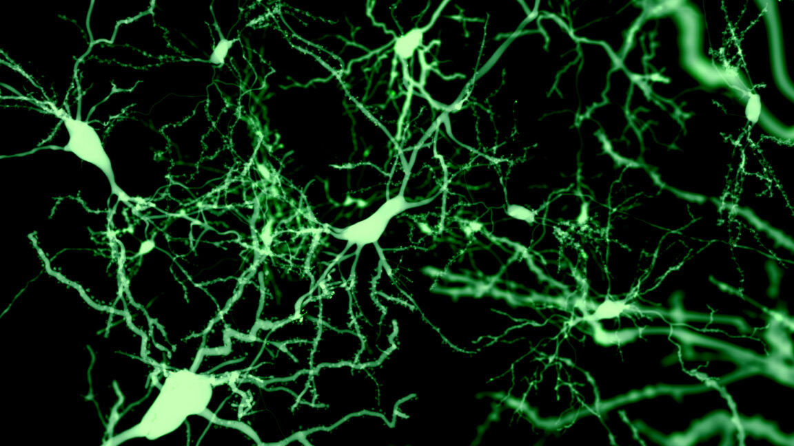
Renata Batista-Brito's team spent years developing a strategy to label these cells by detecting when both genes were active simultaneously. Funding agencies repeatedly rejected the project as too risky.
“I wrote a bunch of grants on these projects that were always rejected because it was too high-risk.”
Massive branching across the brain
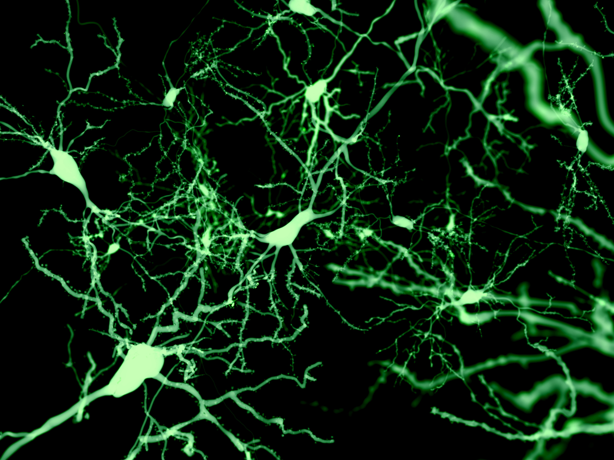
When Batista-Brito finally imaged the Sst-Chodl neurons, she found just two or three cell bodies but massive tree-like branching throughout the visual cortex. Each cell sends connections to areas controlling touch, hearing, memory, navigation, and movement.
“Despite that, there was massive, massive arborization all over the visual cortex, like I never saw with any other neuron.”
They fire during sleep and rest
The team imaged 111 Sst-Chodl neurons while tracking mice's sleep states. Ninety-five of them lit up during slow-wave sleep and quiet wakefulness, but fell silent during running and REM sleep.
Breaking the normal sleep pattern
During deep sleep, the cortex normally alternates between active and silent states. Sst-Chodl neurons break this pattern by firing even more intensely as other neurons quiet down, then skipping the rebound that other cells show.
“Their activity is even higher at the termination of the UP state.”
Controlling the rhythm, not the rate
Using optogenetics to activate Sst-Chodl neurons increased delta waves and tightened the timing of neural firing, but didn't change overall firing rates. The cells coordinate when neurons fire together, not how many fire.
“They change just the coordination, whether the neurons fire together or not.”
One frequency, any stimulus

Remarkably, stimulating Sst-Chodl neurons with different frequencies all produced the same sleep-like oscillation. The cells have intrinsic properties that lock onto a single frequency regardless of input.
“There's something about the intrinsic properties of these cells that, once they trigger, they go on this one frequency.”
Activating them puts mice to sleep
When researchers stimulated Sst-Chodl neurons in freely moving mice, the animals fell asleep faster, slept more during waking hours, and retreated to their nests during the day. The effect was striking even though only a fraction of these rare cells were activated.
“We could have them sleep more during the time that they are awake than they usually sleep during the day.”
Possible sensors of sleep pressure
The researchers propose that Sst-Chodl neurons might sense homeostatic sleep pressure, the accumulated fatigue that forces sleep. Studies show these cells become most active after sleep deprivation.
The mystery of what activates them
The team still doesn't know what turns on Sst-Chodl neurons. They suspect inputs from the hypothalamus and thalamus may trigger sleep-specific activation, which they plan to investigate in the prefrontal cortex.
Conserved from salamanders to humans
Sst-Chodl neurons exist across species from salamanders to humans, suggesting they could be a key target for understanding sleep disorders in psychiatric illnesses. The findings may have broad implications for treating sleep disruption.
The one thing
A tiny population of brain cells called Sst-Chodl neurons, making up just one percent of inhibitory neurons in the cortex, can trigger sleep by synchronizing neural activity across the entire brain.